|
Hoof AnatomyDetails within the Hoof Capsule
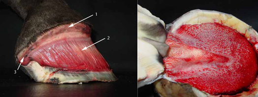
Teaching detailed hoof anatomy is a challenge, but I feel it is important. I have found that understanding the structures has helped me make better choices when trimming. When I began natural trimming, I knew all the parts, but it was a very linear knowledge. I like finding ways to photograph the structures so that their relative positions to each other is understood. I am trying to show the three dimensional aspect to these structures. There is much hidden within the hoof capsule. Here is the first of many layers. Internal Hoof Anatomy
On the left, I have removed the hoof capsule and exposed the 1) coronary band 2) sensitive lamina and 3) the heel bulb. In the next picture, the sole has been removed exposing the papillae that cover the entire solar surface of the hoof.
Hoof Anatomy of the Coronary Band and Coronary Groove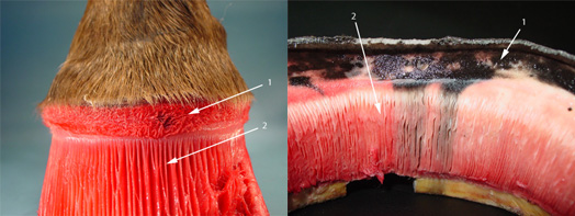
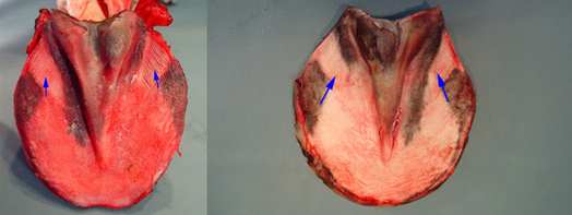
The structures remaining on the leg are the 1) coronary band and 2)sensitive lamina. On the hoof side, you find 1) the coronary groove and 2)the insensitive lamina. I see the black at the coronary groove and soles on many of hooves I take apart. In some hooves, it looks like it is necrotic or dying tissue and in others it appears to be pigmentation. Without proper equipment and histological techniques, it is impossible to know for sure. The stripes going down the insensitive lamina looked very much like necrotic tissue in this particular hoof. I have seen this only one other time and did not get a photo of it. I suspect that sometimes abscesses may move down and do not always track up the foot to the coronary band. I also suspect that small abscess looking marks that are seen growing down the hoof wall are in fact small abscesses that originated in the coronary band. I think of them similar to a pimple. They do not cause lameness because they are small and do not build up the pressure that happens with abscesses encased in the hoof capsule. These are merely my theories and suspicions. Hoof Anatomy of the Sole
The blue arrows are pointing to the sensitive and insensitive lamina on the bars. This hoof was from a young, very flat footed horse in training. The sole was very thin and it did appear that these dark areas were bruises or necrotic tissue. It is hard to make too many assumptions when looking at cadaver feet. I merely see a point in time. Anatomy of the Frog
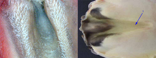
The frog is covered in papillae. These papillae project into small cavities. Some of these small holes are indicated with the blue arrow. Coronary Band and Coronary Groove
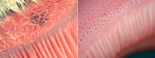
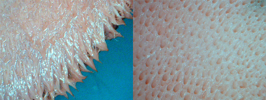
Here is a close up of the papillae on the coronary band and the cavities they project into and the lamina below the coronary band. These papillae supply most of the nutrition to the hoof wall and play a major part in its growth. They produce the pigmented portion of the hoof wall. Papillae on the Sole
The sole corium is covered in papillae that project into the horny tissue of the sole. These papillae are the connection between the corium and the horny tissue of the sole. They supply nutrition and grow the sole of the hoof.
The first of many layers of hoof anatomy.
Return from Hoof Anatomy to Homepage
|

