Clear Equine Distal Limb Anatomy
Tendons and Ligaments Between the Knee and Fetlock

The equine distal limb
Visit my other website, Anatomy-of-the-Equine, to see more anatomy of the lower leg.
There are three bones between the knee and fetlock joints
There are only three bones in this region. In the young horse, they are separate, but often the splint bones fuse to the cannon bone.
- The Cannon bone
- Two Splint bones
- The Suspensory Ligament attaches here
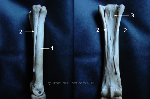
The suspensory ligament attaches behind the knee between the splint bones.
Tendons and Ligaments Revealed
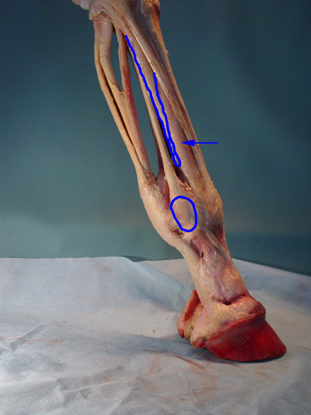
The arrow is marking the cannon bone.
The splint bone has been outlined, and the sesamoid bone has been circled.
The suspensory ligament attaches between the splint bones, then branches out and attaches to the sesamoids. From the sesamoids, it wraps around the front of the pastern and attaches to the coffin bone.
Different Perspective
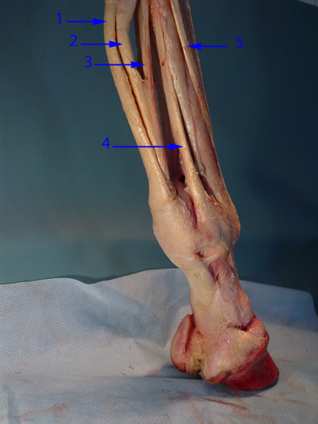
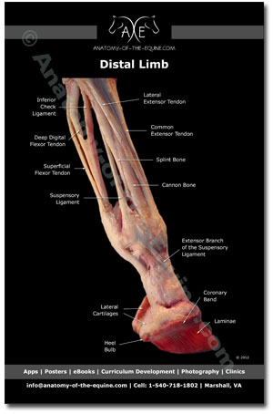
- Superficial Flexor Tendon (SFT)
- Deep Digital Flexor Tendon (DDFT)
- Inferior Check Ligament
- Suspensory Ligament
- Lateral Extensor Tendon
With practice, the different tendons and ligaments can be palpated. Palpation can be a very useful tool to identify regions of pain in the lower leg.
I have stripped away the fascia (connective tissue), so the distinct tendons are ligaments are easy to see, but normally they are bundled together.
Here is a really good video showing the tendons and ligaments
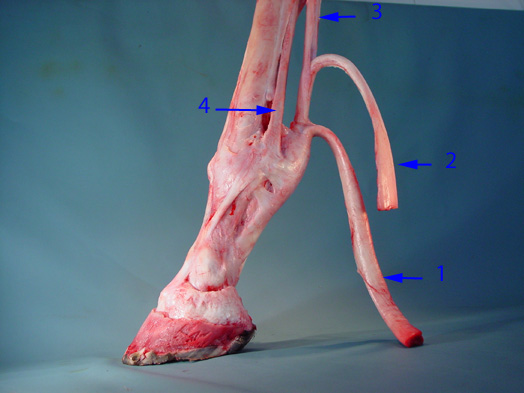
Here is the same information presented in a different way. Hopefully, this helps the viewer piece together the information.
The two flexor tendons have been cut. The inferior check ligament attaches at the back of the knee and attaches to the deep digital flexor tendon.
- Superficial Flexor Tendon
- Deep Digital Flexor Tendon
- Inferior Check Ligament
- Suspensory Ligament
Educational Anatomy Books
New Distal Limb Pocket Guide:
Click here to find out more.
Return from Equine Distal Limb Anatomy to Iron Free Hoof Homepage